My research
From a general point of view, I am interested in the interactions between complex and biological systems and the magnetic field.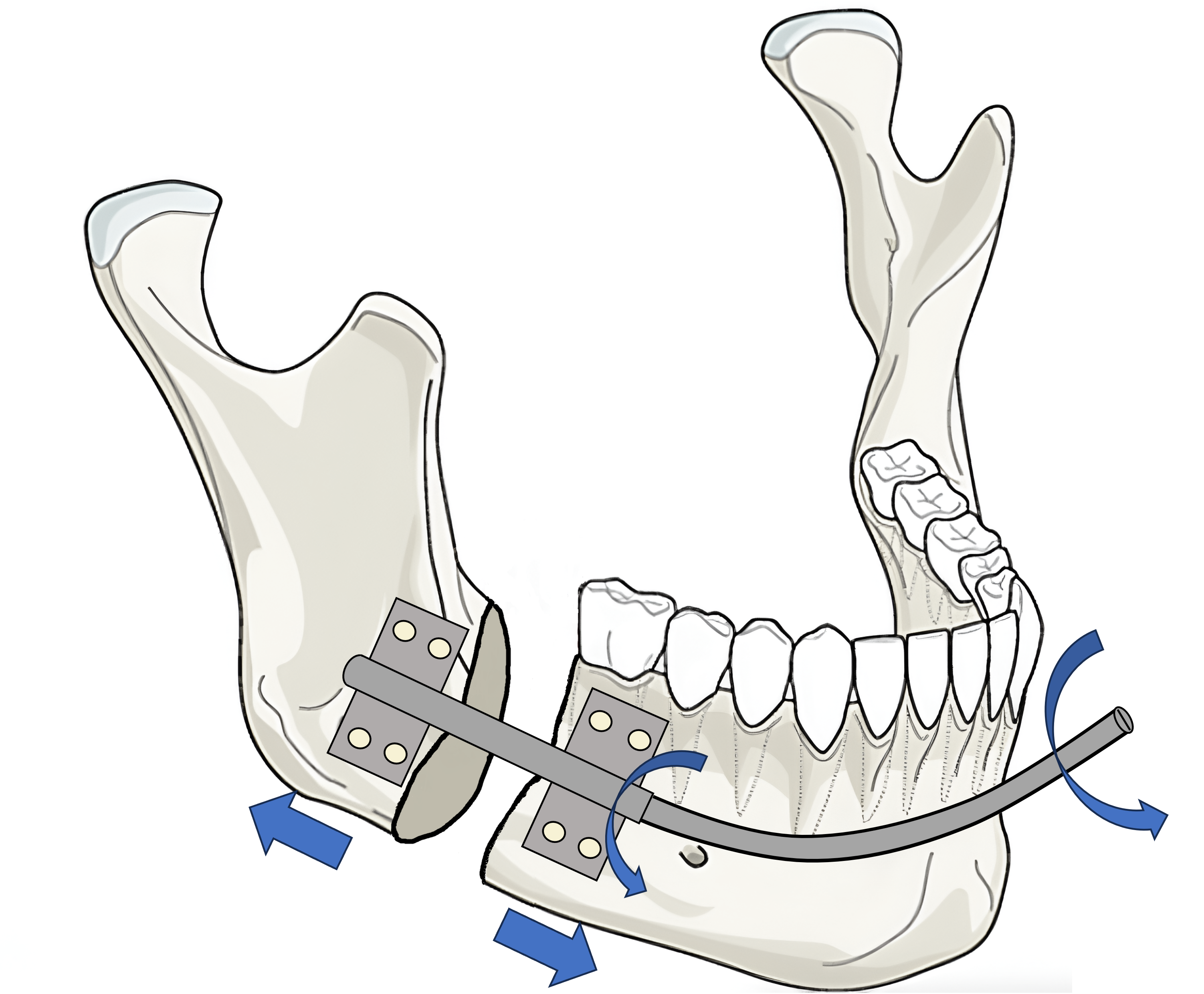
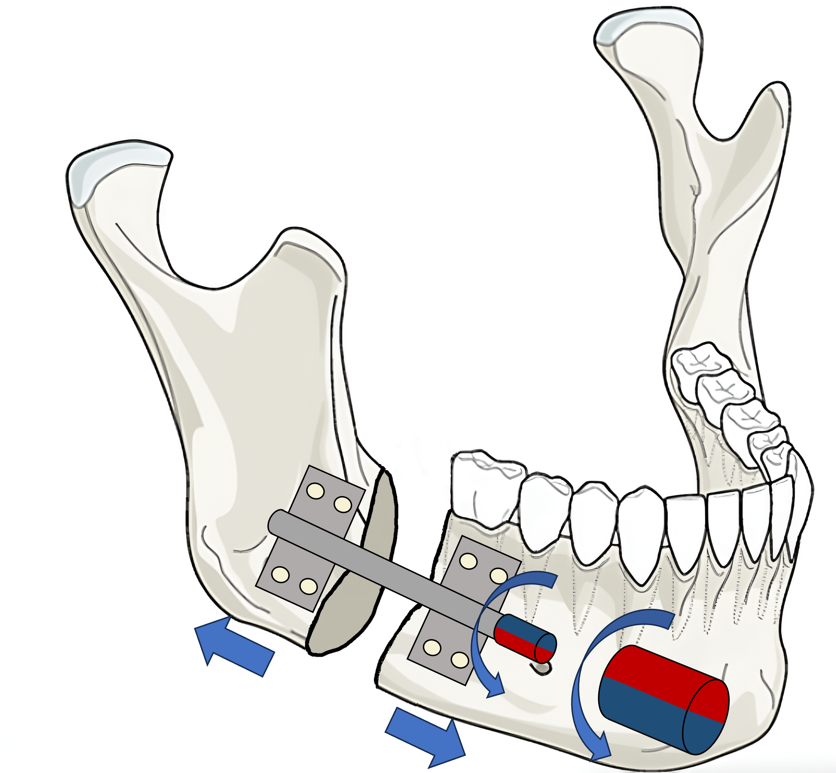 .
.Schematic of a classic distractor with the transcutaneous rod (left), DOGMA magnetic distractor (right).
DOGMA This project consists of developing a prototype of an implantable medical device - a mandibular distractor - which uses the torque transmitted between magnets to activate bone lengthening. Indeed, in certain medical situations, particularly for congenital facial malformations in children, it is necessary to perform bone lengthening by a process called: osteogenic distraction. The surgical operation consists of an osteotomy of the bone to be lengthened, followed by the installation of a subperiosteal distractor. The distractor is composed of 2 plates fixed to the bone on either side of the fracture. These two plates are connected by an endless rod, the activation of which by an externalized rod allows them to be separated, and consequently the separation of the two bone fragments. During the distraction (1 month), the rod which allows the lengthening is a source of complications and suffering. To free ourselves from this rod, we have developed a magnetically activated distractor (DOGMA). The objective of this project is to carry out a clinical study on this new medical device. In this implant, the rod is replaced by a magnet whose rotation, prescribed remotely by an external magnetic activator, causes the bone to lengthen. Currently, the team has produced prototypes of functional mandibular distractors in accordance with regulatory standards as well as designed “smart” external activators, also in accordance with regulatory standards. The activator-distraction assembly was validated on cadaveric subjects at the end of September 2021. The transfer of this technology to a third-party operator began in 2024 for clinical trials to be carried out in 2026-2027.
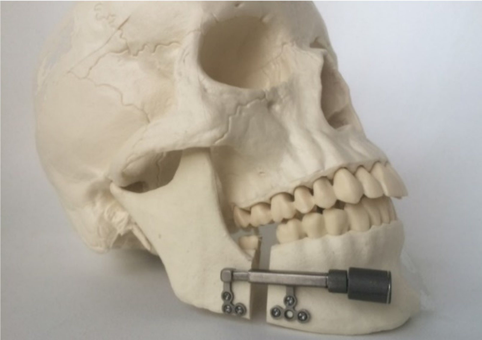
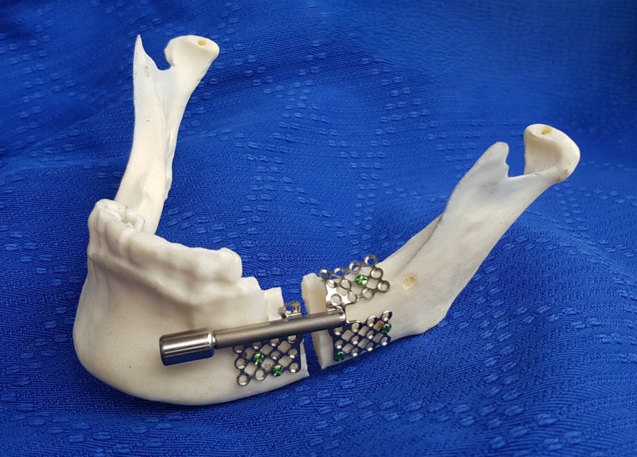
SIMFACE This project focuses on the production of high-fidelity replicas of the face for surgical practice. The objective of this project is to produce high-fidelity replicas, by multi-material additive manufacturing, of biological tissues mechanically representative of the efforts involved in certain surgeries or traumas of the face. During a displaced fracture, the surgical procedure consists of fixing plates on either side of the fracture line ensuring contact and compression between the 2 pieces of bone, thus allowing osteosynthesis. In order to optimize care for trauma and repair of the face observed in the context of the defense health service, we propose to design high-fidelity replicas of the face [1]. This will require reproducing, on the one hand, the mechanical properties of bones stressed in rupture and on the other hand reproducing the particular mechanical properties of biological tissues from materials available in 3D printing. Until 2024, we are focused on this last part by producing composite materials in 3D printing in order to obtain the properties of different soft tissues from living things. These materials are composed of a flexible matrix in which a rigid central fiber is immersed, the geometry of which is optimized to reproduce the target biological tissue. Since then, we have demonstrated the possibility of using a central fiber in a Bézier curve, and validated its use integrated into a flexible matrix.

Composite materials integrating a rigid Bézier fiber in a flexible matrix (V. Serantoni)
However, the central fiber has a characteristic size of the order of a millimeter, which makes the replica incompatible with the morphological constraints of the face while altering the sensations to the touch. It is therefore now necessary to seek to miniaturize the whole. To do this, it is necessary to use a new 3D printing technology, called Melt electrowriting, which makes it possible to produce fibers with a diameter of the order of a micrometer. This will allow us to produce composites reproducing the mechanical properties of the target biological tissues (skin, periosteum, calvaria, etc.) while retaining, on the one hand, a sensation to the touch close to real tissues, and on the other hand, dimensions compatible with the morphological constraints of the face.
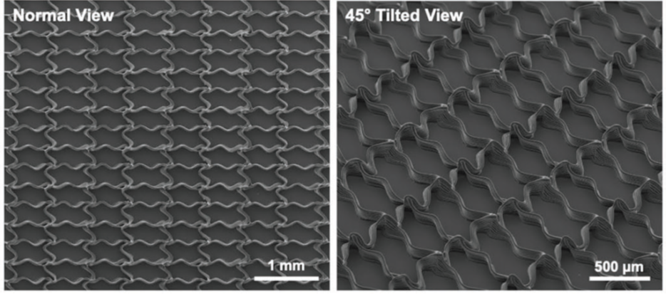
Example of two-dimensional fibers produced with a Melt electrowriting device
MAGBONE This project focuses on the influence of the magnetic field on bone healing. Due to the configuration of the surgery, the internal magnet of the DOGMA device, integrated into the implant, remains close to a fracture line during the entire procedure (4 to 5 months). Its magnetic field could therefore have a direct influence on the bone remodeling process. If, from a regulatory point of view, a static magnetic field below 2 Tesla is considered harmless (Directive 2013/35/EU), adequate data for a proper risk assessment of static magnetic fields are almost completely lacking. Some recent articles have observed that a static magnetic field could have a beneficial influence on the bone remodeling process. The main objective of the MAGBONE project is to improve knowledge on the influence of a static magnetic field on bone remodeling. This is extremely interesting in the context of new medical devices based on permanent magnets, such as the in-house developed mandibular magnetic distractor. The biological aspects will be studied by exposing primary cultures of bone cells (osteoblasts, osteoclasts) to different configurations of magnetic fields (intensity, gradient, orientation). In parallel, we will study the possibility of introducing magnetic particles into the biological environment. Indeed, the latter are rather considered from a therapeutic angle for the treatment of tumors or drug delivery. The presence of these particles combined with external magnets could optimize the interaction of the magnetic field with the biological environment and therefore increase its impact on bone remodeling.

MECADECELL This project aims to understand the impact of bone delcullarization processes on the mechanical properties of bone. The reconstruction of substance losses is a booming surgery that brings together many disciplines. Even more so today, in the era of "appearance", reconstruction with the aim of giving the patient a face that he accepts and with functionality similar to the previous state, is a real challenge for surgeons. The latter use different types of tissues that can be autologous vascularized (free flap, pedicled flap) or not (allograft), or non-vascularized (skin graft, tissue bank). These techniques can be used alone, or in combination and make it possible to provide skin, muscle, and/or bone tissues, in order to recreate the missing volumes. Paradoxically, their realization can involve a considerable loss of substance at the level of the harvesting area, or cause significant complications for the patient: infection, fracture, disunion, necrosis, limited functional results. This is why the use of allogeneic tissues must find its place. Indeed, allografts are already used in maxillofacial surgery and continue to evolve. It is in this context that the first facial allograft was carried out in 2005 by the team of Professors Devauchelle, Testelin and Lengelé. These vascularized allografts make it possible to consider new reconstructions, larger and more complex, but at the cost of heavy and morbid immunosuppression. Long-term immunosuppressants can compromise the graft itself but also the life of the patient with the risk of severe infections, organ failure or cancers. To overcome the rejection of the recipient's immune system, tissue engineering has developed techniques called graft decellularization in order to destroy all cells of the immune system. Thus, the decellularized allogeneic graft can be integrated into the donor while avoiding the usual immunogenic complications, and thus regaining the advantages of an autograft. It is then possible to apply this type of protocol to deceased donors in order to create banks of decellularized flaps.

Paired femurs:
native on the left versus decellularized on the right (U.
Heller).
However, current decellularization techniques are based on bath systems that are ineffective for decellularizing complete bones. Recently, a graft decellularization protocol by perfusion was set up by Professor Lengelé's team in Brussels. This process uses the natural pathways of bones to apply decellularization products, making it possible to produce decellularized complete bones. The first results demonstrated the effectiveness of the protocol. In addition, studies funded by the Gueules Cassées association (U. Heller) were conducted on porcine models to compare the biomechanical properties of decellularized pig forearm bones with native forearm bones. The results demonstrated that the mechanical properties were not significantly altered by decellularization. These initial results are encouraging with a view to using decellularized bone grafts in maxillofacial reconstruction. A study of the characterization of the mechanical properties of grafts after implantation is underway.
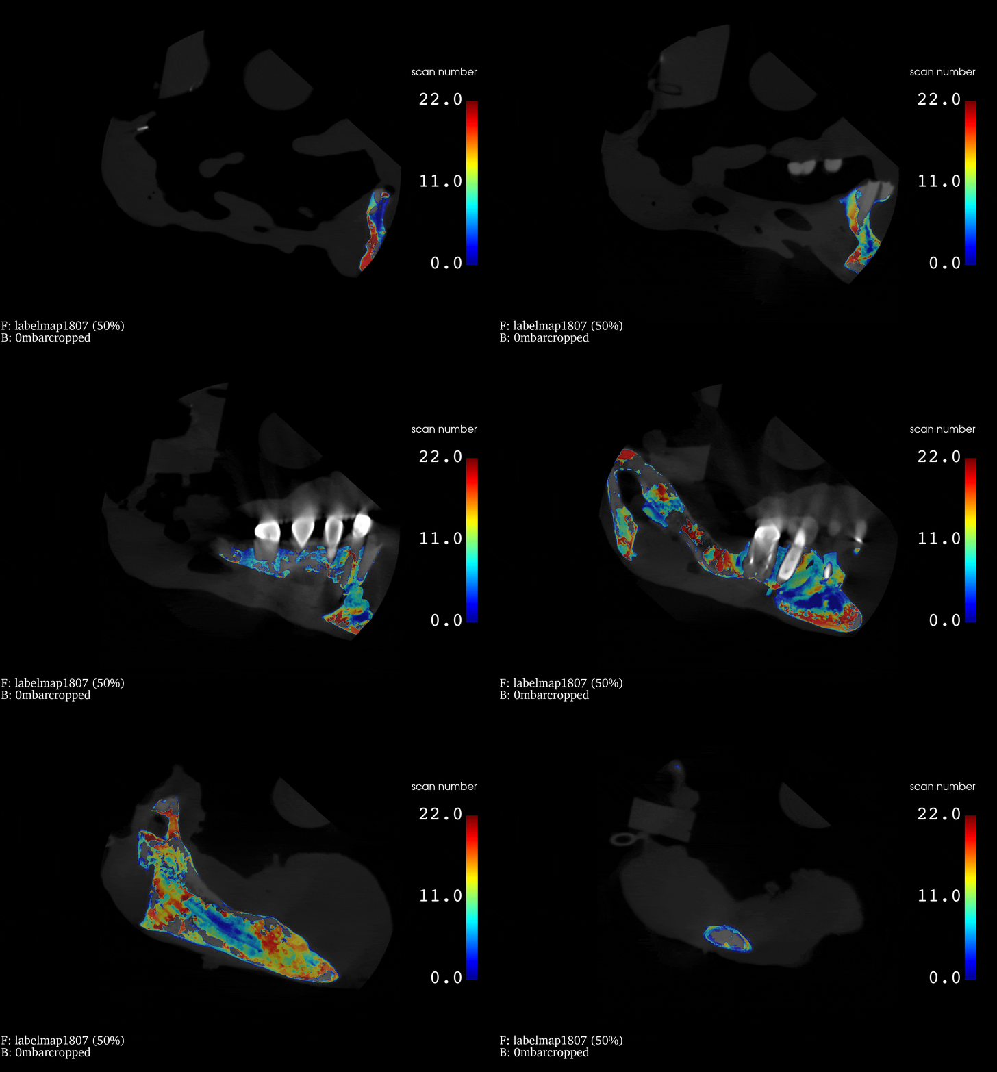
Our research project thus focuses on vascularized bone decellularized by perfusion and more particularly on the mandibular bone. Indeed, in the panel of reconstruction that maxillofacial surgery must take care of, for traumatology, cancerology or the after-effects of radiotherapy (osteoradionecrosis), the mandible is often the first affected, due to its anatomical but also vascular vulnerability. It is as essential, both aesthetically and functionally, as its reconstruction is complex: its interactions are numerous and its geometry convoluted [27]. Obtaining vascularized mandibular bone grafts on demand would therefore be particularly interesting. But its structural complexity and its vascular precariousness contraindicate the identical application of protocols developed on simpler bones, such as the femur.
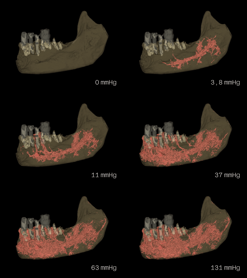
Irrigated territories of the middle mandible as a function of the pressure imposed (C. Serra).
This is why we studied the vascular anatomy of the mandible and its hemodynamic resistances in detail, in order to best export the protocols of decellularization by perfusion. To do this, using a process combining imaging techniques and controlled pressure flow forcing, we experimentally identified the mandibular blood network, and extracted the pressures necessary for its irrigation and the associated pressure losses. Using the results obtained, a specific protocol for decellularization of the mandible by perfusion will be established and tested. This study was carried out using human mandibles taken from anatomical subjects and characterized in the laboratory.
FORMER PROJECTS From a more "classical" mechanical point of view, I am interested in the interactions between complex systems and the magnetic field, whether initially to create electromagnetic forcing systems (pump) or for damping systems (non-linear energy sink, damper with tuned linear or non-linear stiffness, etc.).
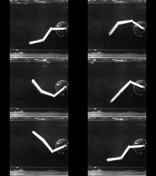
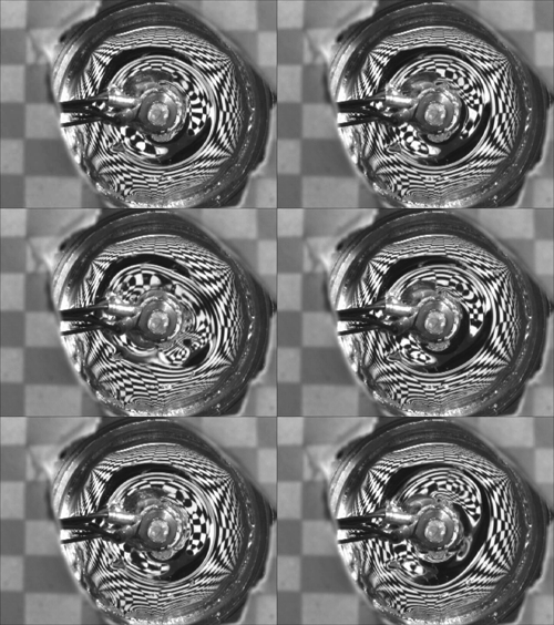
Finally, another of my research themes revolves around magnetohydrodynamics. One of the objectives of this project is to improve our understanding of the physics of conductive fluid flows, in particular the influence of the Lorentz force. To do this, we will work on a Taylor-Couette type model geometry using an experimental device developed at the CEA, in which it is the only force to set the fluid in motion.
